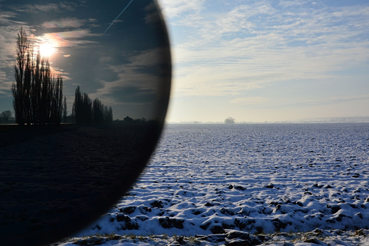

Your eyes are truly remarkable organs. They’re the windows to your world, helping you navigate daily life in countless ways. From picking out the ripest fruit at the grocery store to admiring a breathtaking sunset, your eyes are constantly at work. They let you see your partner’s smile, create stunning works of art, and even visualize the future of our planet. But have you ever wondered how these incredible organs actually function? The anatomy of the eye is an intricate collection of parts, and we’re here to help you see how all the pieces work together to create the miracle of sight.
Warning Signs You Need New Glasses: Don’t Wait
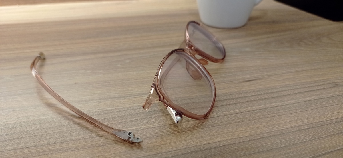
For most of us, we want our glasses to go the distance. They look great, they were a decent investment,